About

veterinary online

New Post

Rss

Sunday, November 25, 2012
veterinary online-The Anatomy of a Factory Farmed Turkey

veterinary online-The Anatomy of a Factory Farmed Turkey

Domesticated turkey


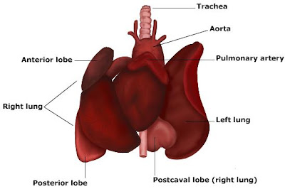
Anatomy of turkey


The turkey was Benjamin Franklin's choice for the United States's national bird. The noble fowl was a favored food of Native Americans. When Europeans arrived, they made it one of only two domestic birds native to the Americas—the Muscovy duck shares the distinction.

Yet by the early 20th century, wild turkeys no longer roamed over much of their traditional range. They had been wiped out by hunting and the disappearance of their favored woodland habitat.

Wild turkeys typically forage on forest floors, but can also be found in grasslands and swamps. They feed on nuts, seeds, fruits, insects, and salamanders.

Wild turkey reintroduction programs began in the 1940s, and the birds were relocated to areas where populations had been decimated but woodlands were recovering. Such efforts worked so well that wild turkeys now live in areas where they may not have occurred when Europeans first reached the Americas. Today, flocks are also found in Hawaii, Europe, and New Zealand.

turkey
Only male turkeys display the ruffled feathers, fanlike tail, bare head, and bright beard commonly associated with these birds. They also gobble with a distinctive sound that can be heard a mile (a kilometer and a half) away.

Females lay 4 to 17 eggs, and feed their chicks after they hatch—but only for a few days. Young turkeys quickly learn to fend for themselves as part of mother/child flocks that can include dozens of animals. Males take no role in the care of young turkeys.

Domestic turkeys have white-tipped tails because they are the descendants of a Mexican subspecies that was taken to Europe for domestication in the early 16th century. The feature distinguishes them from most modern wild turkeys, though captive diet, lifestyle, and breeding have caused other physical discrepancies.
wild turkey

Thursday, November 22, 2012
veterinary online-Interesting facts about Cats

veterinary online-Interesting facts about Cats

In England, a black cat brings good luck and the tail black cat heals sty on the eye, if to put it to the century. For the treatment of warts is the right tail tricolor cat.
 Ancient Vikings also considered the cat sacred. Norwegian Forest Cat mentioned among pets, which the Vikings held on board ships. Similar they trot: thick frill, "brushes" on ears and long hind legs. The first data are dated 1599 year.
Specialists University of Lyon (France) decided to calculate how much the world of home cats. It turned out that they are on the planet 400 million. Most cats in the United States. 
However, the palm give Australia where for every 10 residents accounted for 9 cats. On the Asian continent, first in Indonesia, there are more than 30 million fluffy cat, but in Europe - in France, whose inhabitants are under his tutelage 8 million cats. However, there are countries, such as Peru, Gabon, and some others where domestic cat almost never.
Man respects these magnificent animals. In Japan, for example, at the gates of houses standing figures of cats - a symbol of home and comfort. In Russia, according to tradition, the threshold of a new home first crosses the cat.
As you know, nature had brought humans and animals very different periods of life. Meanwhile, youth, maturity and old age are inherent in all living beings.Naturally, admission to a particular age varies depending on the species. Yes, a month kitten is in the same stage of development as a human baby 5-6 months old. The six-month cat "contemporary" seventh graders (14 years) and fourteen - semidesyatidvuhletneho old.
Cats have one thing in common with giraffes and camels - they "pacers": originally raised both right front and hind legs, and then at the same time - left. In addition, cats - the only animals that do not rely on walking on foot pads, and claws.
cat was in King Charles I, and he believed that without him happiness no issue. He loved her, that she had appointed to guard individual ... but worth overlooked, the cat died. A few days later King Charles was beheaded.
There are many people who are not just in love with these charming tenants, but also connect with them many years of life. Familiar to us from the book of Alexandre Dumas 'The Three Musketeers' insidious personality Cardinal Richelieu was in the palace 12 cats living in his room. To them he showed a lot more good feelings than to people around him. Ernest Hemingway, the famous writer had in his house 150 cats! A world chess champion Alekhine had a live mascot. Favorite Siamese cat chosi (Chess) sat in the hall to his wife's lap during the match Alekhine - Lasker, at a tournament in 1934 in Zurich. Alekhine often descended from the stage in the auditorium to pat his favorite cat.
Traces "mystical" cats are reflected in the tales-remember majestic figure Kota Baiyun - "sitting on a pole, pobyvayuschyy entire nation, whistle Enduring dream and tell a story," Kotofeicha that won the Snake and taught men to make fire. Pushkin Cat Scientist is not so simple: the image of Dubai - a symbol of the World Tree, or Tree of Life, a gold chain - symbol path, (by the way, confused - the word "way"), on which day and night this pundit goes. Correlation with regal goddess of secret knowledge - Isis.Children else know Cat Thief-cat with velvet belly, other significant members of the feline tribe. Story Battle Cat from Dragon into a game of cat and mouse, where Côte always emerges victorious. 
The Russian Old Believers in the early XVIII century cat acted as satirical images of Peter I. When in Egypt entrenched Islam, cats still continued to respect, at least, more than dogs, declared the Koran "unclean animals". And all because personally Prophet Muhammad loved cats - one asleep on his robe, and he had to go to spread Islam. Then he cut the floor where the cat asleep - is not to disturb the sleeping animal.Later, Mohammed, among other titles, also known as the father of cats.
Separately on cat eye: it is a very useful "tool" - the shape and size of the pupil in antiquity could with sufficient accuracy to determine the times of the day. Later invented the photo and began to use the cat eye as Exposure meter.Every self-respecting photographer, except bulky apparatus on a tripod, dragged on his shoulders as cumbersome, fed, full of dignity cat (preferably black). And before the cat had the honor to rise on the shoulders of sorcerers and magicians. 
The Egyptians believed that the soul of the deceased housewife after death hiding in the body of a cat. During a fire, the Egyptians originally carried a cat, and then the stuff. 

As you know, cats are not slaughtered in the fall even from a great height. Why? This question interested professor at Washington University Huayna Whitney. After examining the circumstances of the 132 successful downs animals, he found that cats helps so-called "parachute effect": their legs are extended, and the body expands, reducing the rate of fall. With a minimum height same cat used, primarily, the elasticity of their paws.

All cats and cats - great and people need animals. It is not for nothing that doctors have found that the presence in the house cat or dog brings a person even with infarct zone. Pat cat and decrease blood pressure, silent indignation ... * Back in the 30's, Dr. Joseph Vanco Ryan founded at Duke University (CA), the world's first laboratory parapsychology. As a result of extensive research scientist acknowledged that cats possess paranormal abilities such as foresight and telepathy. Simply put, they can advance to feel approaching danger and at great distances to learn about trouble or death of the owner. Later, other researchers - Nobel Prize winner Niko Tinbergen Dutchman and his colleague Robert Morris - will open in cats ability to use more and psychokinesis (moving objects nonphysical ways), and clairvoyance (getting information on some inanimate objects.) More than half a century, these cat "psi" widely studied in Europe, America and the former USSR.


are owners of cats, leaving a huge legacy of his four-pupils. So, once there was a curious incident described in the book tamer cats Yu Kuklachova "My Favorite Cats." He writes: "In Switzerland there are cats that have their own bank account. Rich housewife put on the account of these animals part of his fortune. And recently, the cats had first exchanged their equity. Royal Society for the Protection of Birds has fined them two pounds "for the systematic killing of birds." A check is signed to care for cats, because bank employees refused to take the form of prints of his paws tailed depositors. " * They say that the invention of iodine assistance had a cat that accidentally overturned flask with different liquids, spilling them. When connected together, the liquid formed a great way as iodine, which is used worldwide for lubrication wounds.
In Japan, in the city of Kagoshima, is the temple of cats. Built it in memory of the seven cats absolutely certain that a commander in 1600, he took with him to war. Cats were soldiers Hours: by expanding or shrinking cat pupil Japanese were able to determine the time. Now this temple visit most watchmakers ... * In ancient Siam, that Thailand is the princess kept cats. When Siamese princess went swimming, then they put their rings on the tail of these cats.
 Since all Siamese cats a bad character and curved tail tip. This legend. And about Siamese cats say they guarded the treasures of Buddhist temples in the jungle, they were the only friends are sitting in prison unnecessary Siamese princes. There is another legend - supposedly Siamese cat in Siam had never lived. And called them so simply because it is the King of Siam gave these cats their favorite British consul. 
Whatever it was, the French magazine "Life of Animals" unearthed for the first time in Europe Siamese cats appeared in London in 1884 hodu.Pohozhee

Wednesday, November 21, 2012
veterinary online-heart, Study of the cardiovascular system of animals

veterinary online-heart, Study of the cardiovascular system of animals

heart, Study of the cardiovascular system of animals

heart

In the study of the cardiovascular system, you must follow a specific sequence: first, examine the area of ​​the location of the heart by inspection and palpation, then make percussion and auscultation body after investigating pulse, if necessary, and use special techniques (electrocardiography, radiography, functional methods, etc. etc.). Establish also vibrate the chest: a burly and long-haired animals, they can not be detected.
heart, Study of the cardiovascular system of animals

veterinary online-Research Respiratory animals

veterinary online-Research Respiratory animals

Research Respiratory animals

Respiratory diseases are widespread among animals, especially young animals. In their study primarily set respiratory rate for 1 min, type, rhythm and symmetry, presence or absence of wheezing and coughing. At rest in adult animals breathing frequency for 1 min is: cattle 12 - 30, a horse 8 - 16, in sheep and goats 16 - 30, in pigs 15 - 20 in dogs 14 - 24, cat 20 -30.
Sunday, November 11, 2012
veterinary online-Veterinary nursing animals

veterinary online-Veterinary nursing animals


Veterinary oncology - the science that studies the anatomical and topographical structure toe and hoof solipeds and kopytets artiodactyls animals, individual characteristics, biomechanics hooves (kopytets), shoeing, research methods, causes of disease, pathogenesis, clinical signs, diagnosis, differential diagnosis, treatment and preventive measures.
Before we treat animals is necessary to examine structural features and functions of the finger and hoof solipeds and kopytets in artiodactyls animals, correct and incorrect statement limbs and its influence on the shape of hooves (kopytets) learn to recognize hoof deformity (kopytets) crack and sternoschisis.
The most common in the hoof (kopytets) is a disease in corolla, crumb, arrows and tissues vault mezhkopyttsevoy slit.
Wounds in the corolla, vaults mezhkopyttsevoy cracks and crumb have the character killed or ushyblenno-torn, almost always positive and usually horses are called "serifs". In artiodactyls wounds can be mezhkopyttsevoy of the corolla, which is well developed mezhpaltsevaya loose fiber.
Wounds are clogged, split and torn. They can be applied by animals (hoof or horseshoe other limbs).This is facilitated by incorrect setting of limbs, improper shoeing, fatigue animal movement on slippery road in deep snow. Wounds can be deposited various metal and other sharp objects.
Superficial wounds can heal without complications. When the skin is damaged bases corolla or conducting layer of the epidermis in some degree disturbed rohoobrazovanyya process. If injured at one point repeating it wound becomes an ulcer. With deep infected wounds corolla may develop abscess, purulent inflammation ungulate joint.
Infected wounds to heel in solipeds may be complicated by necrosis or cartilage development myakishnoho perihondralnoy phlegmon.
In cattle these wounds inflicted by mezhkopyttsevoy slit prone phlegmon and necrosis mezhkopyttsevaya connection.Wounds can have jagged edges, sometimes in the form of flaps. Horses need careful study, as the place injury can cover the hair.
Skin wounds are not usually accompanied by lameness, which appears as a symptom already developing inflammation.
With deep wounds there is a strong pain corolla, when the inflammatory process develops swelling, increased local temperature hoof lameness and there are possible complications of the subcutaneous tissue and other tissues (abscess corolla, necrosis myakishnyh cartilage, suppurative arthritis ungulate joints, etc.).
Superficial wounds with timely treatment usually heal.
In the absence of treatment and distribution of infection can be serious complications developing abscess process. This unfavorable prognosis.
Therefore we must systematically examine hooves and hoof, especially in traumatic injuries and provide immediate assistance.With deep infected wounds and damaged tissues hlubzhelezhaschyh less favorable prognosis in advanced cases - poor.
First aid is smearing wounds and adjacent areas corolla 5% alcohol solution of iodine removal of wool in the corolla over him. The wound was thoroughly washed with soap and antiseptic solutions or make lizolovuyu bath draining wound and surrounding skin with sterile cloth. Secondary grease a 5% solution of iodine above the corolla make circular anesthesia with 1% procaine-it antybyotykovoy solution is removed from the wound of foreign particles, if necessary, dead tissue excised.
The wound was copiously sprinkled complex powder and put moderate pressure bandage soaked yodirovannm alcohol at a dilution of 1:1000 or liniment Vishnevsky. With minor pain reaction and weakly pronounced lameness dressing change on the 3rd day, and balsamic dressing with ointment - to 4-5th day.
A very important condition for the treatment of wounds in the corolla - prevention of pressure horns on the wound surface, liquidation pus pus pockets disclosure. To relieve pressure on its horns stonshuyut coronal edge horny hoof capsule without exposing nipples and leaves skin foundations. Removed only the top layer of horn. Only when detachment hoofed horn in the corolla, when there is a pocket of pus and pus should remove all otsloivshiesya horn.
When excessive growth of granulation appropriate to apply pressure bandage. Pathologically growing horn knife carefully cut ungulates or remove rasp.If the inflammatory process in the corolla is accompanied by a strong painful response and significant disorder limb advisable to use hot tubs. they cause congestion actively contribute to leaching of toxic products, rejection of necrotic tissue, cleaning and reducing pain.
Treatment of gunshot wounds with injury horny capsule and ungulate bones without involvement ungulate joint after careful removal of damaged tissue, using iodoform-ether (1: 10) and dihtyarnym bandages or complex powder usually ends in recovery.
The basis of prevention of injuries and skin corolla in mezhkopyttsevoy gap is proper care of animals.Avoid prolonged race animals in winter on ice and snow. Winter to shoe horses spike only wounded and on the outer branches of horseshoes. We must avoid sudden stops and sedimentation horse back. In some cases where you can not eliminate the notch in the rim, which may be in the wrong setting limbs, apply special nohavki that should be fixed in the tie belt with felt lining.stab wound in the sole of hoof kopytets and arrows often have the character of penetrating wounds.They can be shallow and deep stab, stab.
Such wounds are usually inflicted contaminated sharp objects, fragments of iron nails, glass, wood chips, etc. Often these items may fall into a bed of sawdust. In some cases, they can be the horses in advance to the lapel detached and moved horseshoes. Shallow stab wounds in the area of ​​the front of the foot are less dangerous, because in this area there is subcutaneous tissue (affected base skin). With a deep wound may damage ungulates (kopyttsevidnoy) bone and cast its thin edges.
When wound uninfected subjects and timely treatment can quickly stop the inflammatory response. The most profound adverse infected stab wound. Depending on the injury or hands and soles of the depth of penetration wounds object in the inflammatory process may be involved in the subcutaneous layer of the crumb, arrows and develop abscess.
With deep puncture wounds in animals marked lameness, it does not rely on the affected limb. There have been increasing local temperature, increased pulse finger arteries, characterized knocking pulse.On palpation animal reacts painfully, the presence of a foreign body sticking out above the surface of the foot or hands, can easily
identify the cause of this reaction. In other cases where the foreign body is deeply hidden in the soles or switch grooves need special study. In identifying the object localization wounds after careful preparation, under local anesthesia make its removal. In animals which a foreign body was removed to enter the treatment room its penetration detected after clearing hoof or hooves as spavshehosya, blackened in the lumen of the channel.
When puncture wounds of the foot and hands washed, clearing hoof drew foreign body under local anesthesia. In the event that an animal with splitted wound received the treatment without foreign body in the wound after thorough cleaning well disinfect the wound channel to lubricate the damaged area of ​​5% alcohol solution of iodine, remove nonviable tissue from the wound channel and pipette or syringe injected into it 5%-ing alcoholic solution of iodine and put tar bandage. Animals must put in a clean room with fresh litter. If the wound is superficial, and limp the next day is not enhanced bandage change on 2-3rd day.

veterinary online-Ophthalmology animals

veterinary online-Ophthalmology animals

Ophthalmology animals
Eye Wounds may be superficial and cross-cutting. First aid is to clean the wound from contamination, hair removal and treatment with 1% solution of green fodder. Around the wound for analgesia, enter a 1-2% solution of novocaine. Apply intramuscular antibiotics. Treatment of large-sized wounds operative.After suturing wear protective collar (special circle or plastic bucket).
Inflammation of the eyelids (blepharitis) is accompanied by redness and thickening of the edges of the eyelids, eyelashes appearance at the base of scales and crusts and morning. Eyelashes fall out, the edges of the eyelids strongly thicken. This leads to constant tearing and twisting rumen. Treatment is aimed at removing the cause of blepharitis. Edges century degreased alcohol-ether (1: 1). In the conjunctival sac injected 1%-ing syntomytsynovuyu, 1 -2%-ing oletetrynovuyu, 10%-ing metiluratsilovuyu, 0.5%-ing hentamitsynovoyu emulsion. Good results give 10% probability calendula ointment and daily greasing 1% strength diamond greens. In blepharitis also used betnazol, hydrocortisone acetate, sofradeks intended veterinarian.
In blepharitis caused by Demodex mites, subcutaneously (depending on body weight) administered 0,2-1 ml ivomek.
structure of Eye

Monday, November 5, 2012
no image

veterinary online-Diseases and broken bones of animals

Diseases and broken bones of animals

Causes disease and bone fractures may be closed and open mechanical damage, acute purulent inflammation is localized near the bone.
Peryostyt - inflammation of the periosteum. Acute peryostytah skin in the affected area is treated with a 5% alcohol solution of iodine injected hydrocortisone on 0.25 -0.5% solution of novocaine in the first 24 hours apply cooling bandages. With 2 days prescribed alcohol warming compresses or bandages alcohol, and 3-4-x make massage rezorbyruyuschyh liniment or ointment and heat wrap the affected area. An effective method is physiotherapy daily use paraffin or ozokerytovyh dressings. If pererazdrazhenyya skin rubbing on the area better put a bandage with liniment Vishnevsky.
In introducing peryostytah to education subperiosteal abscess conduct comprehensive treatment. A good therapeutic effect make-novocaine blockade antybyotykovoy with the introduction of antibiotics, hydrocortisone, followed by applying spirtokamfornye or spirtoihtiolovye dressing or hot compress and a course of intensive antibiotic prescribed sulfanilamide preparations.
subperiosteal abscess burst, necrotic tissue and bone sequestration removed and conduct thorough curettage of the affected bone, then wipe the surface of the skin swab moistened with alcohol, ether, sprinkle one with complex antiseptic powder and bandage with liniment Vishnevsky.
In the treatment of chronic periostitis use tools that cause acute inflammation and resorption of fibrous connective tissue in the affected area. When fibrous and formed Osyfikuyuchyy peryostyte conduct spot cautery followed by rubbing ointments that aggravate inflammation. For resorption of fibrous tissue successfully used pirogenal or tissue therapy. These tools are useful and when osteophytes and exostoses (bone formation). Effective use of ultrasound.
osteitis - inflammation of bone - as an independent disease is rare. Typically, the inflammatory process involved almost all the elements of bone: periosteum, bone, endosteum and bone marrow.
In osteitis fault area is saturated alcoholic solution of iodine. To reduce exudation in the first day prescribed cold and oppressive. Further, as the disappearance of acute inflammation to accelerate resorption of fluid using heat treatments in a warming compresses and condenses under osteitis - paraffin and ozokeritotherapy for deep heating.
Necrosis - necrosis of bone that occurs when purulent | inflammatory processes in the different layers of bone (purulent peryostyte, osteomyelitis), mechanical injuries (bruises, I concussions, fractures), physical (frostbite, burns) and chemical influences. These factors lead to thrombosis of vessels that nourish the bone. Depending on the degree of damage can occur necrosis complete or total when omertvevaet whole bone or most of it, and incomplete or partial when the affected part of the bone. In addition, the localization of bone necrosis can be superficial (cortical) nekrotyzuyetsya outer surface of bone, and the central (deep) which omertvevayut its deep layers. Treatment should be prompt and sent to remove necrotic areas. Later prescribe a course of intensive antibiotic therapy by intraosseous injections.
Caries - limited grained molecular breakdown of bone formation on the surface of the bone defect (bone ulcers). Treatment is mainly surgical, aimed at autopsy fistulous passages, removing dead tissue and to ensure a good flow of fluid. Then apply antiseptic drugs mouthwashes.
Osteomyelitis - inflammation of the bone marrow, endosteum, periosteum and compact substance.There aseptic osteomyelitis and infectious. The clinical course they are acute and chronic. Factor is bone injury, exhaustion, cooling, vitamin deficiency and severe contagious disease that reduces bone marrow resistance and resistance infection. The main causes of suppurative osteomyelitis are open fractures, penetrating to the bone infected wounds, festering Periostitis.
In the initial stage of osteomyelitis intraosseous (in the bone marrow canal) injected Novocain, antibiotics in N. Yelanskomu against previously provided lrotivosepticheskoy therapy. Needle with mandrenom or sleeve with stylet left in the bone marrow cavity and vbintovyvayut in a bandage in order to perform these injections of 1 - 2 times a day. In the presence of subperiosteal abscesses coming just as in peryostytah.
The formation intermuscular phlegmon them reveal, remove dead tissue, bone sequesters, stop bleeding, use complex aseptic powders and treats as if phlegmon. After easing acute inflammation conduct trepanation bones reveal bone marrow canal, remove diseased bone marrow, bone sequesters, carefully conducted curettage, wash the affected area of ​​hydrogen peroxide, then alcohol-ether for 5 - 7 minutes and fill the cavity of the complex antiseptic powder containing iodoform and antibiotics. The surface of the wound is closed with a towel and put uhlehipsom bespodkladochnoy plaster cast or bandage with liniment Vishnevsky. Simultaneously, a course of intensive antibiotic therapy, actively using sulfanilamide preparations, vitamins, crystalline trypsin. Highly is intraosseous introduction to the damage area 5 - 10% solution yodoformennoho ether in a dose of 3 - 5 ml of 1 day a week. For treatment use 3 - 5 injections.
Fractures. Anyway fractures tubular or flat bones there is a gap of muscles, fascia, blood vessels, nerves, organs, and in open fractures damaged skin and other soft tissue. Fractures can be congenital or acquired, closed and open, complete and incomplete.
Types of fractures and osteosynthesis for fractures shown in Fig.
Antibiotic for dogs at perelome
broken bones
Rice. Types of fractures animal and - boundary fracture b - breakdown, in - complete fracture
veterinary online-bone(Humerus)

veterinary online-bone(Humerus)


Humerus

Definition

Bones together, asymmetrical forms the skeleton of the arm. Long bone diaphysis 1, 2 epiphyses.

Situation

It is structured:
  • upwards, inwards and backwards with the glenoid cavity of the scapula in his head.
  • bottom with the forearm by means of the distal humerus:
  • down and out with the humeral capitulum radius by.
  • downwards and inwards with the ulna by the humeral trochlea.

Orientation

The spherical surface at the top and inside the edge projecting furthest.

General anatomical shape

  • Triangular bone cut to the middle 1/3 of its shaft, he describes three sides and three edges:
    • posterior, anterolateral, anteromedial face.
    • anterior, medial, lateral edge.
  • Head 30 mm radius representing third of a sphere.
  • Neck-shaft angle of 130 °.
  • Humeral lopsided 30 to 45 ° forward.
  • Retroversion angle of the humeral head 20 ° to the distal humerus.
  • Angle of the head relative to the horizontal: 45 °.

Palpable landmarks

  • greater tuberosity
  • lesser tubercle
  • lateral and medial epicondyle
  • furrow inter tuberosity.

Relations

  • Axillary nerve (C5-C6), radial (C6-C7-C8-Th1), ulnar (C8-Th1)
  • Axillary brachial artery and deep
  • Long head of biceps tendon and ligament Transverse humeral.

Clinical

  • Fracture at the neck: axillary nerve injury (more shoulder abduction above 8 °)
  • Fracture in middle 1/3 of the diaphysis: radial nerve injury (hand gooseneck) and lesion of the brachial artery deep.
  • Fracture of the medial epicondyle: ulnar nerve injury (ulnar claw)
  • Synovial inflammation of the tendon of the long head of the biceps: tenosynovitis
  • Dislocation of the glenohumeral: control of the axillary nerve to perfor

bone-Humerus


veterinary online: Scapula bone

veterinary online: Scapula bone

Scapula bone
 Scapula bone

Definition

Bones together, flat and asymmetric. It forms with the clavicle shoulder girdle.

Situation

It is located on the posterior surface of the rib cage, pressed against the ribs 7 and 2. 
It is structured:
  • with the clavicle upwards and inwards by the articular surface of the acromion.
  • top outside forward by the glenoid with the humeral head.
  • There is a syssarcose between the anterior and posterior of the scapula of the rib cage.
Scapula

veterinary online- bones

veterinary online- bones

BONES

Bone structure and blood supply 
The osteology is the study of bones. All the bones that form the skeleton is the framework of the body, serves to support soft tissue and plays a protective role against certain viscera. Bone is living in perpetual redesign, even in adults
Description bones :There are three types of bone:
Long bones: the length is greater than the width and thickness.
A middle part, the body or shaft of the bone.
Two ends: the epiphyses.
Or large flat bones: the length and width are greater than the thickness.
Both sides.
Edges.
Short bones: the length, width and thickness are approximately equal.

Bone structure

Bone structure

1)Bone tissue.
2)The bone cells .
3)The osteoblasts : bone forming cells: secrete collagen, a protein substance necessary for ossification.
4)The osteocytes : star-shaped cells with thin and long extensions combine the cells together These are mature bone cells.
5)The osteoclast : cells that destroy bone while osteoblasts rebuild parallel.
6)The osteogenic cells: which are formed from all tissues.
7)The bone ground substance : ossein and minerals.
8)The varieties of bone tissue 
9)The compact fabric : the basic substance is dense. It forms the outer layer of the bone, it covers the spongy tissue.
10)The spongy tissue : the basic substance is less dense, rich in fat and connective tissue cells in red marrow where blood cells form.

Structure of different bones

1)The shaft : made ​​of compact bone tissue
2)The cortical width of the hollow cylinder formed by the compact bone tissue.
3)The cavity or spinal canal : cavity dug into the shaft in the middle of the cylinder compact. 
4)The yellow bone marrow : mainly greasy tissue filled the medullary canal.
5)The epiphyses : formed of spongy tissue filled with red bone marrow. They have surfaces which articular cartilage can be linked to each other.
6)The metaphyses : segment of bone between the epiphysis and the diaphysis.
7)The periosteum : fibro-elastic membrane that envelops the entire bone except at joint surfaces and cartilage.
8)It contains many blood vessels and bone provides a portion of its feeder vessels.

Development and growth of bones

The development of bone

Bone development

The development of bone osteogenesis. Bone can be formed by two different processes:
Endochondral ossification or cartilage ossification : each bone is preceded by a cartilaginous. This draft was then invaded by blood vessels. There is destruction of cartilage cells by connective tissue cells which then produce bone. This ossification begins during intrauterine life. There for long bone diaphyseal ossification point and one or more epiphyseal ossification.
Ossification fibrous or ossification membrane : the cells of the connective tissue of the embryo form a blank conjunctiva future bone and they transform directly into bone cells or osteoblasts which will develop the final bone.
The increase in length : it is through the growth plate. The diaphysis of the bone under the action of cartilage increases in length until early adulthood. The growth rate is controlled by a hormone (STH somatotrophyne or secreted by the pituitary gland). The thyroid has an important role on growth.

The increase in thickness : it is done through the periosteum. The periosteum, the deep layer develops layers of bone. At the same time, there is an increase in the medullary canal. The bone material is destroyed by osteoclasts. The medullary bone preserves considerable robustness while lightening.


Copyright © 2014 veterinary online All Right Reserved